Optical Imaging Center Erlangen (OICE)

The OICE offers and coordinates a direct access to modern microscopy technology. The involvement of the OICE within the CRC 1181 allows scientists to obtain the possibility to get a more detailed insight into the molecular mechanisms and processes from a macroscopic to nanoscopic scale.

Technics and methods

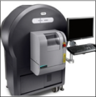
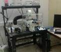
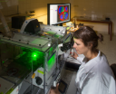
Modalities IVIS Spectrum & µCT Intravital Microscope Spinning Disc LSM Confocal LSM STED / RESOLFT iSCAT / STORM
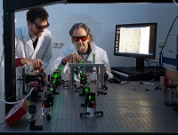
Quelle: Ralf Palmisano / OICE Quelle: Ralf Palmisano / OICE Quelle: Ralf Palmisano / OICE Quelle: Ralf Palmisano / OICE Bild5 Friedrich Alexander Universität Erlangen Nürnberg Optical Imaging Center Wissenschaftler Laser Labor Wissenschaftler Ralf Palmisano Tristan Nowak
Technics
  • Molecular and anatomical views of complex biological systems
  • high resolution (up to 4.5 micrometer voxel size)
  • Multimodal 3D Imaging with parallel tomographic imaging of fluoresence and luminesence signals
  • Longitudinal studies to  monitoring disease progression in vivo
  • Compensation possibility for motion artefacts (i.g. of breath and cardiac action)
  • High resolution multiphoton imaging
  • Spectra Physics Insight DeepSee Dual
    (680 – 1300 nm) Laser – high penetration depth
  • 2 GaAsP NDDs in objective halter
  • Investigation of different tissue in vivo
  • high penetration depth
  • High resolution confocal imaging
  • Longtudial living cell microscopy
  • normoxic and hypoxischen
    environmental factors
  • Fast mulit-camera -system


  • Multichannel fluorescence
  • High resolution confocal imaging
  • 8 laser excitation wave lengths (405 ~ 647 nm)
  • 6 detector units (3 PMTs, 2 HyDs, 1 TMP)
  • Extremly fast point scanner ( max. 8.000 Hz)
  • Normoxic living cell microscopy
  • Super high resolution fluorescence microscopy
  • Fixed samples and living cell microscopy (normoxic)
  • 3 canal STED
  • 3 APD detectors
  • 3D STED compedent
  • RESOLFT longtudial living cell microscopy
  • Super high resolution imaging
  • No labelling needed
  • single-molecule imaging
  • single-molecule tracking
Resolution  macro 1µm micro 200nm  micro 200nm  micro 160nm nano 2D 10 x 10 x 500 nm oder 3D 90 x 90 x 90 nm nano 5nm, Localisation precision < 1 nm

Contakt